Negative staining – TEM sample preparation
For biological objects hard to see
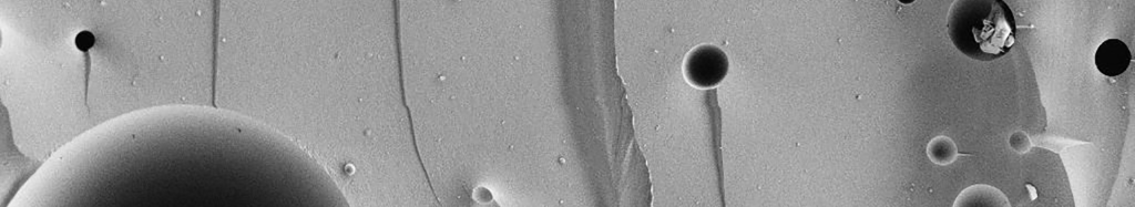
Isolated organelles, individual macromolecules and viruses pose a problem when imaging in a TEM since they do not have high enough contrast (they are not electron-dense).


Negative staining
Biological objects need to be stained in a way that helps to distinguish their edges and internal structure. For this purpose, we use negative staining.
It is the perfect tool when it comes to the size distribution of liposomes, exosomes or polymer nanoparticles. The limitation of this technique is resolution to a maximum of ~18 – 20 Å.
What is negative staining?
The negative staining process begins by placing a drop of your sample onto a TEM mesh grid. The grid is covered with a support film made of an amorphous carbon layer, sometimes supported by a thin layer of polyvinyl film (e.g. Formvar, Pioloform).
After the sample is adsorbed, blotted and washed if necessary, we apply a staining reagent for a very short time (10-40 sec). The stain is then blotted off, air-dried and the grid is ready to be imaged.
The most commonly used negative staining reagents are uranyl acetate and uranyl formate. These stains have a relatively fine grain size (4 – 5 Å) and provide higher resolution images over other stains. Uranyl acetate and formate also act as a fixative, preserving many protein-protein interactions on a millisecond time scale, although the low pH of the stain and its propensity to precipitate at physiological pH may be detrimental to some samples.
How the service works
When you send us your sample, we prepare it and use negative staining, then image it to your specification and within your timescale. For the imaging part see TEM imaging service. If your sample requires centrifugation prior to use, please advise us about the best conditions and centrifuge speed.
How to make sure your sample is prepared in the best way
Fixed or unfixed samples may be used. Samples should be resuspended in a suitable buffer (e.g. 0.1M Sodium Cacodylate, pH 7.0) or in distilled water. It is best not to use phosphate buffer or PBS as they may contaminate the grid with salt residues that have to be washed off after staining resulting in a loss of contrast. Reducing agents, detergents, sucrose, glycerol, and high concentrations of nucleotide should also be avoided as they also affect stain quality.
This may not always be possible so we can adhere your sample (liposomes or similar) to the grid, wash off the residual solution and then carry on negative staining. This reduces formation of buffer-related artefacts and generally improves staining. If we expect buffer artefacts, we can prepare a ‘control’ grid – a buffer-only grid stained exactly the same, to determine if the buffer components are the source of the observed artefacts.
Concentration of your sample
The more concentrated the sample, the better. We only need 10 ul of your sample at most, the minimal volume is 4 ul to place on a TEM grid.
Frequently Asked Questions
We try to avoid this problem by fixing the sample after it has adhered to the grid with glutaraldehyde. The negative stain – uranyl acetate – also helps to prevent the sample from shrinking by fixing it further. Structures that are not rigid, such as large vacuoles mostly formed of water, can collapse inward once dried so the internal shape of these structures should not be considered as it may be an artefact.
The best method for transporting your sample depends on its nature. If your sample needs to be frozen, it should be transported on dry ice. For samples that do not require freezing, chilled transport should be sufficient.
The cost for negative staining preparation for TEM is approximately £90 for up to 15 samples. Additionally, there is a charge of £5 per sample for consumables. Imaging one sample costs around £90, particularly if the sample is highly dense.


