Techniques
- Biological sample preparation
- Immunolabelling
- Cryo sample preparation
- Metal sample preparation
- Sample preparation for SEM
- Time scale of sample preparation
Sample preparation
1. TEM
The first step of preparing the sample is fixation. In general, we use 2.5% glutaraldehyde due to its excellent ability to preserve microstructure but other fixatives or combined approaches can be used, such as using both paraformaldehyde and glutaraldehyde.
Then, processing the tissue by penetrating it and fixing it with osmium tetroxide is the next step. Here, additional procedures that are required are usually implemented here such as immunostaining.
Dehydration is a necessary step as all our microscopes work in vacuum and thus require the sample to be free of water. This is performed in ethanol or acetone,
In a graded series increasing the concentration of the alcohol.
Next, the sample needs to be infiltrated with resin, which when polymerized, creates a hard medium, embedding the sample that can be sectioned.
So called flat-embedding is suitable for thin sliced tissue (usually using vibrotom) 50-150um thick. In this technique tissue slices are embedded between two Aclar films (Agar) and then stuck to an epon block for sectioning. The advantage of this is that only small amount of tissue used, orientation of embedded tissue is easy to know and 40-50-um can be immunolabeled before flat-embedding.
The sections are cut using an ultramicrotome, either thick several microns for light microscopy or ultrathin under 80 nm for electron microscopy.
The last step is contrasting the sections using uranyl acetate and lead citrate.

Cryo sample preparation (Impact freezing and freeze-substitution)
Impact freezing is used to quickly freeze and thus fix samples without significant ice crystal damage. This contrasts with chemical fixation performed at room temperature which takes longer. Impact freezing is mostly used for special purposes (e.g. immunolabelling of antigens with antibodies that are aldehyde-sensitive).
Impact freezing of the sample is followed by freeze-substitution, during which the fixation occurs. Water in the cells is substituted by organic solvent at temperatures between -80 °C and -90° C. Chemical fixatives (e.g. glutaraldehyde, uranyl acetate or osmium tetroxide) are then introduced, resulting in diffusion into cells. Cellular components are immobilized by low temperature, and when substitution medium is warmed up to the temperature that permits chemical cross linking, the fixatives are already dispersed throughout the cells and fixation occurs immediately. Embedding and polymerization are done at low temperatures (between -30 °C and -55 °C).

Immunolabelling
Pre-embedding immunolabelling
Vibrotomed tissueslices ~40um thick are immunolabelled prior flat-embedding. For immunolabelling, tissue slices are fixed with PFA-fixative and permeabilized by Triton-X100. For detection of immunolabelling, we use Fab-fragments of secondary antibodies conjugated with ultra-small gold nanoparticles. The use of small gold particles and Fab-fragments allows efficient diffusion into the tissue. Gold nanoparticles are then enhanced with silver enhancement. Then tissue slices are flat-embedded.
Post-embedding immunolabelling
Cells are fixed with formaldehyde/glutaraldehyde mixture or impact frozen, and then embedded in Lowicryl HM20 resin (TAAB) at cold temperatures using freeze substitution method. Labelling is done on ultrathin plastic sections using secondary antibodies conjugated with 5, 10 or 15 nm gold particles (Aurion) or protein A-conjugated with 5, 10 or 15 nm gold particles (Aurion).

Preparation of metals
Preparation of metals for TEM will involve the preparation of thin 3mm diameter discs, in which a hole is created using the jet-electroplishing technique.
The EM Suite has a range of equipment designed for these preparation methods, including:
- Polaron SC7640 Sputter Coater with Au target
- Edwards Model 306 Carbon CoaterMetalthin Jet-Electropolishing Cell for preparation of metallic TEM specimens
- Metallographic specimen preparation (mounting, polishing etc.)
2. SEM
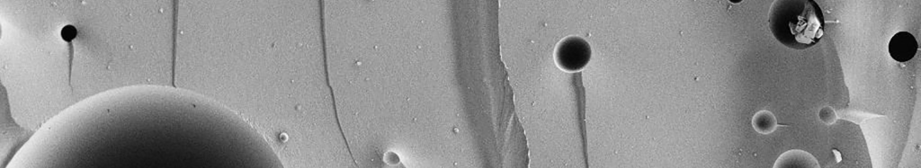
Sample preparation for SEM primarily depends on whether or not the sample is conductive. Additionally, biological samples may need to be dehydrated either using chemical or gas dehydration techniques.

Sample preparation for SEM primarily depends on whether or not the sample is conductive. Additionally, biological samples may need to be dehydrated either using chemical or gas dehydration techniques.

Non-conductive samples may be coated with a conductive film of metal or carbon, in order to render them conductive.
Time scale
Generally, the preparation of samples for imaging by TEM takes at least 2 weeks. If specialized techniques of sectioning or processing are used, this may take longer, from 4 weeks onwards.
Preparation times for SEM samples will vary from minutes to weeks depending on the nature of preparation required. Some samples require very little processing.
Biological samples may require sectioning, dehydration processes which may take days.
Metallurgical samples may require sectioning, mounting, grinding polishing, etching etc. which may take some days.


